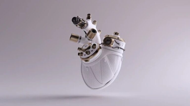
Human tissue bioprinting is still in development, including the ability to 3D print entire human organs, and a team of researchers at Carnegie Mellon University have announced their ability to 3D print full-size components of the heart just like natural ones.
The paper was published in Science, and one of the researchers who conducted this work in his lab says: “We were able to print efficiently functioning parts of the heart, like a heart valve or a small beating ventricle. Using the MRI data of the human heart, we were able to: Accurately reproduce the patient’s anatomical structure, 3D collagen and human heart cells.” He added, “It is important to understand that there are many years of research yet to be conducted, but there is still cause for excitement. We are making real progress towards engineering tissues and functional human organs, and this paper is only one step on the way.” (1) .
And it’s not just the Carnegie Mellon researchers, but other researchers from the University of Minnesota have developed in 3D-printed models of the aortic heart valve and surrounding structures that mimic the exact look and feel of a patient.
Published in Science Advances, the researchers 3D-printed the so-called aortic root, the part closest to and attached to the heart. The aortic root consists of the aortic valve and the openings of the coronary arteries, and contains three layers called leaflets surrounded by a fibrous ring. The model also includes a portion of the left ventricular muscle and the ascending aorta.
But what is the purpose of printing these forms?
“Our goal with these 3D-printed models is to reduce medical risks and complications by providing patient-specific tools to help clinicians understand the precise anatomical structure and mechanical properties of a specific patient’s heart,” says a University of Minnesota Mechanical Engineering researcher.
This model is specifically designed to help doctors with a procedure known as transcatheter aortic valve replacement (TAVR). It is a procedure to treat aortic stenosis by implanting a vital prosthetic valve into the affected original valve (2).
Aortic root models are made using CT, It is a type of imaging that uses special X-ray equipment to make cross-sectional images of the body, thus proportional to the exact shape, and then 3D-printed using specialized silicone-based inks that mechanically match the feel of real heart tissue. Commercial printers on the market today can 3D print, but they use inks that are often too rigid and not suitable for the smoothness of true heart tissue (2,3).
On the other hand, specialized 3D printers at the University of Minnesota were able to simulate the soft tissue components of the model (2).
But the researchers do not see this achievement as the end of the road for these 3D-printed models. Do you think, dear reader, that this printing will stop at this point, but will continue to develop?
Sources :
1. Durham E. 3D printing the human heart [Internet]. : Here
2. Haghiashtiani G, Qiu K, Zhengre Sanchez J, Fuenning Z, Nair P, Ahlberg S et al. : Here
3. CT Scans: MedlinePlus [Internet]. : Here


0 Comments